Featured Articles

Boost Your Website Traffic with High Quality DA/PA 40+ Backlinks
Apr 7, 2023

In today’s competitive world, one must be knowledgeable about the latest online bus...
Oct 12, 2018
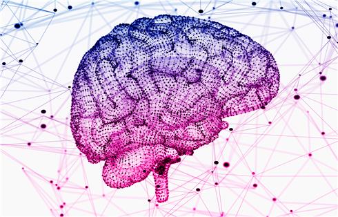
Nervous System Development Overview
The development of a single human fertilized egg into an individual is largely mysterious. In the process, the developmental central nervous system is the most complex. The central nervous system is formed by the ectoderm of the embryo. In the neuroblast stage, the notochord is the central axis of the embryo that runs through the embryo early and induces the undifferentiated ectodermal cells above it to transform into the primordium of the central nervous system. First, the dorsal ectoderm cells above the notochord are elongated and thickened to form a front wide and narrow nerve plate; the edge of the nerve plate is thickened and pleated to form a nerve pleat; the central plate of the nerve plate is concave to form a nerve groove. Then, the nerve pleat moves to the midline of the back and finally closes to form a neural tube. The anterior part of the neural tube develops into a brain, and the posterior part develops into a spinal cord. From this stage, the general characteristics of the brain are determined by the growth and bending of the anterior portion of the neural tube. With the gradual enlargement of the cerebral cortex and the production of cortical folds, a mature brain is formed, and in the early stages of human embryonic development, human fetuses and mammalian fetuses have striking similarities. This is the total neuroanatomical feature of the development of the nervous system.
Nervous System Development Function
The development of the nervous system is especially important for the body. The development of the nervous system mainly includes the following four aspects.
The proliferation of neurons: a primary question about brain development is whether the brain neurons are produced before birth or after birth. Studies on the brains of human newborns have shown that newborns have resemble brain with the adult, except that the axons of brain neurons are not myelinated, but there is already a well-developed brain. Virtually all neurons are produced before birth, although some studies have recently modified this theory. Through the cell labeling study of pregnant embryos, the timing of neuronal production can be clearly shown. The primate cerebral cortex is produced during the first quarter of pregnancy. During the period, all the number of cortical neurons at birth was produced.
Neuronal movement: The cortical-forming neurons are produced in the cell layer near the ventricles of the brain. This cell layer is called the ventricle zone, where the cells divide to form cortical neurons. Neurons and glial cells in the ventricles begin to migrate along a special glial cell, from the ventricle to the cortical surface, and the moving neurons stay on the growing cortical surface for a short time, then move later. Neurons stay in more superficial locations beyond previously moving neurons. Therefore, the cerebral cortex is a layered structure from the inside to the outside. The initially moving neurons are located at the deepest part of the cortex, while the last moving neurons stay in the more superficial position. Because the timing of the production of cortical neurons determines the final position of cortical neurons, all effects on the movement of cortical neurons lead to a deformed cortex. A typical example of human neuron being destroyed during movement is the developmental damage. If a mother abuses alcohol for a long time, the movement of fetal neurons will be severely damaged. As a result, the fetus produces an abnormal cerebral cortex, which inevitably leads to physical and cognitive and emotional defects.
Neuronal Type Decisions: Current neuroanatomical studies all cerebral cortical cells, including neurons and glial cells, which are known to be derived from undifferentiated precursor cells in the ventricles. The precursor cells move to the layers of the cortex by movement. Once the position of the precursor cell is determined, the type of cell will become and the location of the cortex to be stopped will be determined.
Synapse production: Synapses in the brain are produced before birth, but do not reach vertex density until fifteen months after birth. Synapses are produced earlier in the cortical deep layer, but later it also produces in the cortical surface. At the same time as synapses are produced, brain neurons also increase the area of neuronal dendrites, extend axons of neurons, and undergo myelination. Synaptic deletions can also occur at the same time as synapses, which is probably a way to regulate central nervous system connections. It may be to remove excessive or ineffective connections between neurons. Synapses at different locations of cerebral cortex are produced at different times, providing a basis for maturation of different regions of the cerebral cortex at different times. In addition, in the development of the central nervous system, if it is supported by a rich external environment and experience, it may lead to more developed dendrites and axons and more synapses.
Nervous System Development Research Status
Nervous system development is a highly complex and highly coordinated process, and signal transduction plays a decisive role in neural development. Wnt signaling pathway is involved in the regulation of multiple functions of the body. In nervous system development, Wnt signaling pathway is involved in neurogenesis, axon guidance, and dendritic development during neurodevelopment. Once the Wnt signal transduction in this process is abnormal, it will cause neurological development defects and cause many neurological diseases. In specific regions of the mammalian brain, endogenous neural stem cells (NSCs) can differentiate into functional neurons, a process called neurogenesis. The Wnt signaling pathway plays an important role in the development of the nervous system and the development of neural stem cells such as proliferation and differentiation. In Wnt signaling pathway, Wnt3a regulates the proliferation and differentiation of neural stem cells in neurogenesis. Wnt3a activates the canonical Wnt signaling pathway and increases the expression of β-catenin, which can promote the differentiation of neural stem cells into neuronal cells and even inhibit the differentiation of neural stem cells into astrocytes. When Wnt3a is overexpressed in the cerebral cortex of mice, neural progenitor cells induce cortical layers to differentiate into neurons and accumulate at the margin of SVZ/IZ. Conversely, in the middle and late stages of neural stem cell differentiation, the signaling pathway was blocked by the inhibitor of Wnt signaling pathway -DKK1, and neuronal production was inhibited. When the Wnt3a gene was knocked out, the proliferation and differentiation of endothelial stem cells were significantly inhibited. Activation of the upstream Wnt ligand of this signaling pathway or inhibition of GSK-3 protects neurons and promotes differentiation of neural stem cells to reduce damage. Activation of Wnt5a in non-canonical signaling pathways also promotes and regulates the proliferation of NSCs to dopaminergic neurons. In summary, the open signal pathway of the Wnt signaling pathway is involved in neurogenesis, and plays an important role in the differentiation of embryonic and adult neural stem cells and the proliferation of neurons. It plays a role in the development of the nervous system. In the future research, further understanding of the transduction mechanism of Wnt signaling pathway and the inhibition of related diseases will play an important role in the treatment of human-related diseases in the future.
BMP signaling and neurogenesis: Studies have shown that the BMP signaling pathway plays an important regulatory role at all stages of neurogenesis and plays different roles at different stages and at different sites, sometimes even the opposite. It shows function of promotion and inhibition for proliferation, which may be due to the difference in responsiveness of neural stem cells to BMP signals at different stages. During the development of the central nervous system, the early bmp gene is highly expressed on both sides of the nerve plate, thereby exerting a function of defining the region of the neuroectoderm. When the nerve plate is closed to form a neural tube, it is mainly in the dorsal midline, ie, the top plate. Studies have shown that the apical plate is important for the regulation of neural tube closure and proliferation and differentiation of neural stem cells in the neural tube. The BMP protein secreted by the apical plate forms a high to low concentration gradient from the dorsal to the ventral side in the neural tube and plays a key regulatory role in the above process. In addition, the transgenic analysis has revealed that the use of Nestin's neuron-specific enhancer to regulate the expression of BMPRIA in the early stage of mouse neurogenesis to enhance the activation of BMP signaling pathway leads to an increase in the proliferation of neural stem cells and a significant increase in the number. During the chicken embryo neurogenesis, overexpression of constitutively activated BMPRIA at the cerebellum can result in an increase in the number of granulosa cells. These studies have shown that activation of the BMP signaling pathway is essential for neural stem cell proliferation during neurodevelopment. Studies have shown that Wnt at least partially mediates the role of BMP signaling in maintaining neural progenitor cell proliferation. It has also been found that as a responsible gene for BMP signaling, zic1 can inhibit terminal differentiation of neural progenitor cells by inhibiting the expression of the proneural gene math1. The classical mechanism of pluripotency maintenance of neural stem cells is the side-inhibition model mediated by the Notch signaling pathway: the surface of the neonatal neurons begins to express the ligand of Notch, which activates the Notch signaling pathway of adjacent neural stem cells through cell-to-cell contact, thereby initiating neural stem cells to maintain the expression of the gene hes1. Through this mechanism, newborn neurons can effectively inhibit the early differentiation of adjacent neural stem cells and maintain a certain number of neural stem cells.
References:
- Franze K. The mechanical control of nervous system development. Development. 2013, 140(15):3069-3077.
- Alyaa M, Moiz B. Role of Cytokine Signaling during Nervous System Development. International Journal of Molecular Sciences. 2013, 14(7):13931-13957.
- Tam S J, Watts R J. Connecting vascular and nervous system development: angiogenesis and the blood-brain barrier. Annual Review of Neuroscience. 2010, 33(1):379-408.
- Yaron A, Sprinzak D. The cis side of juxtacrine signaling: a new role in the development of the nervous system. Trends in Neurosciences. 2012, 35(4):230-239.
- Nakano H, Murabe N, Amemiya S, et al. Nervous system development of the sea cucumber Stichopus japonicus. Developmental Biology. 2006, 292(1):205-212.
Article source: https://article-realm.com/article/Health-Fitness/Medicine/55510-Nervous-System-Development.html
Comments
Reviews
Most Recent Articles
- Jun 30, 2026 Why Choosing the Right Eye Specialist for Children in Kolkata Matters by Alzbeta Berka
- May 20, 2026 ABA Billing Companies | Top ABA Billing & Revenue Cycle Experts by julyjack
- May 18, 2026 Global Orthodontic Headgear Market Analysis, Trends, Report, 2034 by Dipak Straits
- Mar 5, 2026 Early-Stage Clinical Trial Mistakes Sponsors Overlook by Guest
- Dec 30, 2025 Kinase Inhibitors for Cancer Treatment Market Insights 2025–2033 by Dr. Vikas Singhal
Most Viewed Articles
- 6843 hits What You Need to Know About the Prostate Gland and Its Symptoms by Brentconroy
- 582 hits Discover The Best Professional Fitness Coach In Newcastle And Wollongong by Thegorilla Pit
- 527 hits Should I Choose Physiotherapy Over Other Treatment Options by Tom Cartwright
- 525 hits Comprehensive Review of Designs for Health Supplements: Berberine-Evail, GI Revive, and ArthroSoothe by Ella Smith
- 510 hits For what reason would it be advisable for me I be utilizing Whey Protein by dhamisingh
Popular Articles
In today’s competitive world, one must be knowledgeable about the latest online business that works effectively through seo services....
81007 Views

Walmart is being sued by a customer alleging racial discrimination. The customer who has filed a lawsuit against the retailer claims that it...
50438 Views
Are you caught in between seo companies introduced by a friend, researched by you, or advertised by a particular site? If that is...
37043 Views

Facebook, the best and most used social app in the world, has all the social features you need. However, one feature is missing. You cannot chat...
23354 Views
If you have an idea for a new product, you can start by performing a patent search. This will help you decide whether your idea could become the...
14578 Views

Moving becomes easy when you have the right moving accessories. These moving accessories help secure and protect your item by ensuring that no harm...
12617 Views

A lot of us look forward to the result of moving and not the process itself. It is pretty typical behavior, though. As modern people, many things...
11969 Views

Building a custom home is an exciting adventure. It’s your chance to bring your vision to life and create an area that sincerely displays...
11740 Views

Moving from one state, city, or even to a whole different county, is something that is either dictated by choice or circumstance. This is because,...
11420 Views

A membrane contactor is a device that enables the transfer of components between two immiscible phases, typically a gas and a liquid, through a...
10434 Views
Statistics
| Members | |
|---|---|
| Members: | 16750 |
| Publishing | |
|---|---|
| Articles: | 78,497 |
| Categories: | 202 |
| Online | |
|---|---|
| Active Users: | 2783 |
| Members: | 16 |
| Guests: | 2767 |
| Bots: | 8937 |
| Visits last 24h (live): | 25912 |
| Visits last 24h (bots): | 40671 |


